First Step of Radiotherapy: Positioning with Radiotherapy Immobilization Mask
Radiotherapy is a treatment for tumors that uses high-energy X-rays or other types of radiation to kill tumor cells. Radiation beams are generated by radiotherapy machines, among which linear accelerators are the most commonly used. The rays penetrate healthy human tissues to reach tumors. Radiation acts like a double-edged sword: while destroying tumor cells, it may also injure adjacent normal organs. Modern precision radiotherapy equipment and techniques deliver higher radiation doses to tumors while reducing exposure to normal tissues.
Precise positioning serves as the first step of precision radiotherapy. The primary goals of positioning are to acquire CT imaging data under treatment posture, which is applied for target volume delineation and treatment plan design. In addition, the same posture captured during positioning must be reproduced every day once treatment starts. Better posture consistency leads to higher treatment accuracy.
CT simulation positioning is the most widely adopted method at present. Positioning protocols vary for tumors at different anatomical sites, yet the general workflow remains consistent.
Taking head and neck tumors as an example, we introduce the CT simulation positioning process below.
01
Under the guidance of radiotherapy technologists, the patient lies supine on the CT table with a head-neck-shoulder support underneath and a dedicated radiotherapy pillow for head support. The head is slightly tilted backward, and the body is kept as straight as possible. Next, a softened head-neck-shoulder thermoplastic mask, a high-tech shaping material that softens in hot water at approximately 60℃, is placed over the patient’s head, neck and upper chest area.
The thermoplastic mask cures and forms a rigid fixation shell after cooling for 5 to 10 minutes. This process is similar to tailor-making, creating a personalized mould for each patient to deliver reliable immobilization and ensure excellent posture reproducibility.
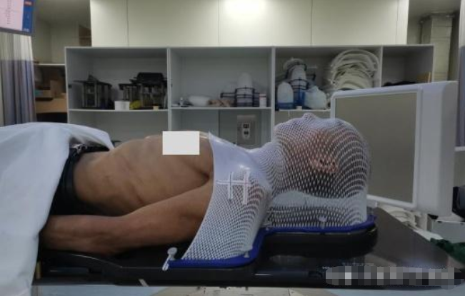
The thermoplastic mask features a perforated design. There is no risk of suffocation even when it covers the entire facial region.
02
Technologists then draw three cross markers aligned with laser lights to define positioning isocenters. Three lead markers with a diameter of 1 mm are attached onto these isocenter points, followed by plain or contrast-enhanced CT scanning.
After removing the mask, technologists also mark the laser-aligned cross lines directly on the patient’s skin. The markers on both the mould and skin form a dual safeguard to maintain posture reproducibility.
This completes the positioning procedure. The patient only needs to wait for radiotherapy plan design before treatment delivery.
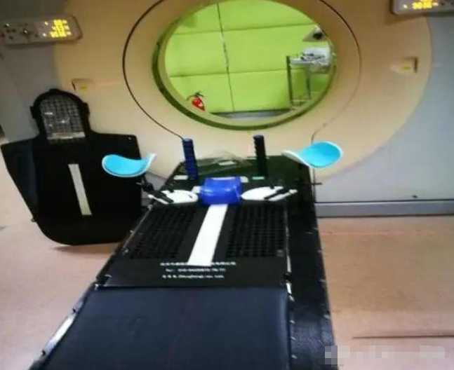
Throughout the whole treatment period, all marker lines on the patient’s skin must be kept intact and should not be scrubbed off at will. Technologists will retouch faded lines after daily treatment sessions.
For breast cancer patients, moulds are generally unnecessary for radiotherapy. Only specialized breast positioning brackets are used, with positioning markers drawn on the skin. Moulds may raise radiation dose to the skin and increase skin adverse reactions.
Although positioning sounds straightforward, it involves numerous details, and patients need to complete a series of preparations before positioning.
For instance: Patients with pelvic tumors need to drink 500–600 ml of water about 40 minutes in advance to maintain adequate bladder filling, empty the rectum as much as possible and stay moderately fasting; Patients with head and neck tumors must remove metal dental appliances before positioning; For lung cancer patients, lesions shift with respiratory motion. Respiratory motion management or 4D-CT positioning is required. This technique acquires CT images across multiple respiratory phases for image fusion and delineates the moving range of the tumor as the treatment target volume.
Therefore, full patient cooperation under technologists’ guidance is critical. Physicians and medical physicists also participate in this process.
maidfirm Thermoplastic Radiotherapy Immobilization Mask
maidfirm provides high-performance thermoplastic radiotherapy immobilization masks tailored for clinical CT simulation and radiotherapy positioning. Made of premium medical-grade polymer raw materials, our positioning masks deliver stable mechanical properties and consistent stretching performance after heating. Available in multiple specifications including head mask, head-neck mask, head-neck-shoulder mask, with various perforation rates to meet different clinical immobilization requirements.
Our mask softens rapidly in 60–70 ℃ warm water, featuring good ductility for seamless fitting on patient contours. Fast cooling and high dimensional stability ensure outstanding daily posture repeatability during fractionated radiotherapy. The breathable perforated structure reduces patient stuffiness and eliminates suffocation concerns. All maidfirm radiotherapy positioning products comply with medical device standards, widely adopted in head & neck, thoracic and other tumor radiotherapy immobilization scenarios, helping clinics achieve precise and safe radiotherapy positioning.